- Gleneagles Medical Centre 6 Napier Road, #09-19, Singapore 258499

We strive to help you manage urogynaecologic symptoms so you can get back to living your best life. Book consultation with Dr Arthur Tseng now!
References:
Samuelsson EC, et al. Signs of genital prolapse in a Swedish
population of women 20 to 59 years of age and possible
related factors. Am J Obstet Gynecol. 1999;180:299-305.
Swift SE. The distribution of pelvic organ support in a
population of female subjects seen for routine gynecologic
health care. Am J Obstet Gynecol. 2000;183:L277-85.
Al-Allard P, Rochette L. The descriptive epidemiology of
hysterectomy, province of Quebec. Ann Epidemiol. 1991;1:541-
9,1981-1988.
Hilton P, Dolan LM. Pathophysiology of urinary incontinence
and pelvic organ prolapse. BJOG. 2004;5-9.
Mant J, et al. Epidemiology of genital prolapse: observations
from the Oxford Family Planning Association study. Br J Obstet
Gynaecol. 1997;104:579-85.
Samuelsson EC, et al. Signs of genital prolapse in a Swedish
population of women 20 to 59 years of age and possible
related factors. Am J Obstet Gynecol. 1999;180:299-305.
Rinne KM, et al. What predisposes young women to genital
prolapse? Eur J Obstet Gynecol Reprod Biol. 1999;84:23-5.
Spence-Jones C, et al. Bowel dysfunction: a pathogenic factor
in uterovaginal prolapse and urinary stress incontinence. Br J
Obstet Gynaecol. 1994;101:147-52.
Jorgensen S, et al. Heavy lifting at work and risk of genital
prolapse and herniated lumbar disc in assistant nurses. Occup
Med. 1994;44:47-9.
Wiskind AK, et al. The incidence of genital prolapse after the
Burch colposuspension. Am J Obstet Gynecol. 1992;167:399-
404.
Holley R, et al. Recurrent pelvic support defects after
sacrospinous ligament fixation for vaginal vault prolapse. J
Am Col Surg. 1995;180:444-8.
Chaikin DC, et al. Predicting the need for anti-incontinence
surgery in continent women undergoing repair of severe
urogenital prolapse. J Urol. 2000;163:531-534.
Barrington JW, Edwards G. Posthysterectomy vault prolapse.
Intl Urogynecol J Pelvic Floor Dysfunc. 2000;11:241-245.
Delancey JO and Hurd WW. Size of the urogenital hiatus in
the levator ani muscles in normal women and women with
pelvic organ prolapse. Obstet Gynecol. 1998; 91:364-8.
Veronikis DF, et al. The incidence of low-pressure urethra as
a function of prolapse-reducing technique in patients with
massive pelvic organ prolapse (maximum descent at all
vaginal sites). American Journal of Obstetrics & Gynecology
1997;177(6):1305-14.
Richardson DA, et al. The effect of uterovaginal prolapse
on urethrovesical pressure dynamics. Am J Obstet Gynecol.
1983;146:901-5.
Romanzi LJ, et al. The effect of genital prolapse on voiding.
Journal of Urology 1999;161(2):581-6.
Versi E, et al. Videourodynamic diagnosis of occult genuine
stress incontinence in patients with anterior vaginal
wall relaxation. Journal of the Society for Gynecologic
Investigation 1998;5(6):327-30.
Suzanne Hagen, et al. Conservative management of pelvic
organ prolapse in women. Editorial Group: Cochrane
Incontinence Group. 21/01/2009.
Pott-Grinstein E, Newcomer JR. Gynecologists’ patterns of
prescribing pessaries. J Reprod Med. 2001;46:205–8.
Handa VL, Jones M. Do pessaries prevent the progression of
pelvic organ prolapse? Int Urogynecol J. 2002;13:349-52.
Brubaker L, et al. Chapter 5 – Pelvic Organ Prolapse.
Incontinence (Editors: Paul Abrams, Linda Cardozo, Saad
Khoury, Alan Wein). 2nd International Consultation on
Incontinence Paris, July 1-3, 2001. 2nd Edition 2002.
Babak, et al. Levator contraction strength and genital hiatus
as risk factors for recurrent pelvic organ prolapse. American
Journal of Obstetrics and Gynecology 2005;192(5):1592-8.
J&J Marketed Vaginal Mesh Implant Without U.S. Approval.
Bloomberg. David Voreacos and Alex Nussbaum – Mar 21,
2012.
Johnson & Johnson’s Ethicon Division to Stop Selling
Gynecare Vaginal Mesh Systems. Chaffin Luhana. New York,
NY (PRWEB) June 06, 2012.
As the more visible chronic diseases become easily manageable, other less visible diseases are coming to the forefront as the baby boomers in our population experience life in their golden years. Pelvic organ prolapse (POP) is one such neglected entity that adversely affects a patient’s quality of life, and can only become more prevalent with time.
The prevalence of prolapse varies from 2-48%. It was estimated that 30% of all hysterectomies for POP occurred in women greater than 50 years old (compared to 7% in the 15-49 year age group). The annual incidence of hysterectomy for prolapse peaked in the 65-69 year age group at around 30 per 10,000 (0.3%).

It is important for women who are experiencing pelvic organ prolapse to seek medical advice.
The pelvic organs are supported by muscles, connective tissue, ligaments and nerves that form a “pelvic floor” in the pelvis. When these structures become weakened or stretched, the organs can slip out of place and cause pelvic organ prolapse. Risk factors for POP include childbirth, ageing, obesity, smoking and chronic coughing or straining.
Different types of Pelvic Organ Prolapse (POP) are defined based on which organ is prolapsing. These include:
A cystocele refers to the descent or bulging of the bladder into the anterior vaginal wall. It occurs when the supporting tissues between the bladder and the vaginal wall weaken or stretch, resulting in a protrusion of the bladder into the vagina.
A rectocele is the descent or protrusion of the rectum into the posterior vaginal wall. It happens when the supportive tissues between the rectum and the vaginal wall weaken or stretch, causing the rectum to push against the vaginal wall.
Uterine prolapse involves the descent or sagging of the uterus into or through the vaginal canal. It occurs when the ligaments and muscles that support the uterus become weak or damaged, causing the uterus to drop down towards or even outside of the vaginal opening.
Vaginal vault prolapse refers to the descent or dropping of the upper portion of the vagina after a hysterectomy. It occurs when the supporting structures that hold the vaginal vault in place become weakened or stretched, leading to the vaginal vault descending into the vaginal canal.
Enterocele is the descent or herniation of the small bowel through a defect in the vaginal apex. It happens when the supporting tissues in the pelvic floor become weakened, allowing the small bowel to push into the vaginal space.
Urethrocele is the descent or bulging of the urethra into the anterior vaginal wall. It occurs when the supporting tissues around the urethra become weak, causing the urethra to protrude into the vagina.
If you experience symptoms such as:
It is important to consult with your healthcare provider. They can perform a physical examination and other tests to determine if you have pelvic organ prolapse.
The diagnosis of pelvic organ prolapse (POP) is typically made through a physical examination and medical history. Your doctor may also use imaging tests, such as an MRI or ultrasound, to further evaluate the extent of the prolapse.
During a physical exam, your doctor will assess the position of your bladder, rectum, uterus and vagina. They may also perform a pelvic floor muscle strength test to evaluate the strength and function of your pelvic floor muscles.
When patients present with POP, assessment for urinary incontinence is required as both conditions are interconnected. There is the entity of occult urodynamic stress incontinence (USI), which has an incidence rate of 23-50%.
Medical conditions that are treatable should be assessed and therapy optimisation instituted, including improving asthma control, correcting constipation, or controlled weight loss. Previous conservative, medical and surgical treatment for POP should be noted as they affect treatment outcomes
Routine urine microscopy and culture is performed to exclude infection, and an ultrasound of the pelvis is useful in excluding gynaecological conditions that require urgent treatment (e.g. ovarian cancer) or impact on prolapse (e.g. large fibroid causing pressure effects). An ultrasound of the upper renal tract can be done for severe POP, as up to one-third of patients may have obstructive uropathic changes.13 Ultrasound can also be used to delineate the pelvic floor to aid in localising defects.
An essential part of assessment would be urodynamic studies, which comprises uroflowmetry, simple filling and voiding cystometry and urethral pressure profilometry. These tests are performed when urinary symptoms are complex and/or there is a need for surgery. Uroflowmetry is a useful screen for possible voiding dysfunction.
Cystometry involves inserting catheters into the bladder and rectum to monitor pressure changes with respect to volume of instilled sterile fluids. It allows for the diagnosis of urodynamic stress incontinence (USI) and occult USI, detrusor overactivity (DO) or mixed urinary incontinence and causes of voiding dysfunction. There is a 3% risk of UTI with urodynamics, hence preventive antibiotics are given immediately after the procedure.
The treatment for POP is dependent on the patient’s age, prolapse severity, underlying medical conditions and childbearing wishes. Generally, once women have prolapse, it is impossible to recover, hence prevention is important. An experienced obstetrician to manage the pregnancy optimally is required. This involves constipation control, monitoring weight increase and estimated foetal weight, and antenatal and post-natal pelvic floor exercise (PFE).
It can also be
a temporising measure for patients
awaiting surgery. The commonest
is a ring pessary, used to support
and relieve prolapse symptoms.
Patients fitted with a pessary will
require regular pelvic examinations
every three to four months unless
they can successfully remove and
insert their pessary themselves.
At
a review, the doctor can remove the pessary, cleanse the vagina and
exclude ulcers before replacing
the pessary with a new one. The
judicious use of topical oestrogens
aids in maintaining mucosal
integrity and should be restricted
to a maximum of three months for
patients with an intact uterus.
In cases of utero-vaginal
prolapse, a vaginal hysterectomy
(VH) can be performed. In cases
of a hyper-elongated cervix, a
Manchester’s operation can remove
the prolapsing cervix whilst
retaining the uterus. In cases of
cystocele and/or rectocele, repair of
the herniation is called an anterior
and posterior colporrhaphy or a
pelvic floor repair (PFR).
In cases
of vault prolapse, a sacrospinous
ligament fixation (SSF) attaches the
vaginal apex to the right ligament
(usually) with non-absorbable
sutures.
The commonest side
effect is short-term pain in the
right gluteal region, which is
relieved by simple analgesics.
In almost all cases, reconstruction
of the disrupted perineal body
(perineorrhaphy) is paramount in
supporting the entire pelvic floor.
The reduction in vaginal aperture
diameter also reduces pressure
transmission forces that predispose
to prolapse. Post-operative advice
is given (i.e. to not carry heavy
loads, to avoid constipation and
to not squat) as these actions may
increase prolapse recurrence.
There are attendant risks with general anaesthesia and they should
be made known to the patient before surgery. There are also surgical complications which include risk of bleeding, infection at wound site and trauma to the bladder or rectum. These risks are low (around 1%) when surgery is done by experienced surgeons.
Atelactatic pneumonia can be prevented by deep breathing exercises and early ambulation. Deep vein thrombosis (DVT) and pulmonary embolus (PE) can be prevented by using TED stockings and pneumatic calf compression devices, early ambulation and low-molecular weight heparin (LMWH) injections.
It was considered a safe and effective procedure, with long-lasting cure rates, no serious complications and mesh erosions rates of 10%-16%. In July 2011, the U.S. Food and Drug Administration (FDA) issued a bulletin stating that individuals with transvaginal mesh kits had complaints of “mesh becoming exposed or protruding out of the vaginal tissue (erosion), pain, infection, bleeding and pain during sexual intercourse, organ perforation from surgical tools used in the mesh placement procedure, and urinary problems”.
Some reports cited the need for additional surgeries or hospitalisation to “treat complications or to remove the mesh”, and the number of complaints between 2008 and 2010 were reported at “1503 adverse event reports associated with mesh used for POP repair”. This prompted a “review of scientific literature published between 1996 and 2010 comparing mesh surgeries to non-mesh surgeries.
The agency review suggests that many patients who undergo transvaginal POP repair with mesh are exposed to additional risks, compared to patients who undergo POP repair with stitches alone. While mesh often corrected anatomy, there was no evidence that mesh provided any greater clinical benefit than non-mesh surgeries.” This triggered certain companies to withdraw prolapse mesh kit products and cease production amidst a flurry of mounting legal complaints. Currently, natural tissue repair appears to be the safest method and has a low complication rate if done by experienced specialists
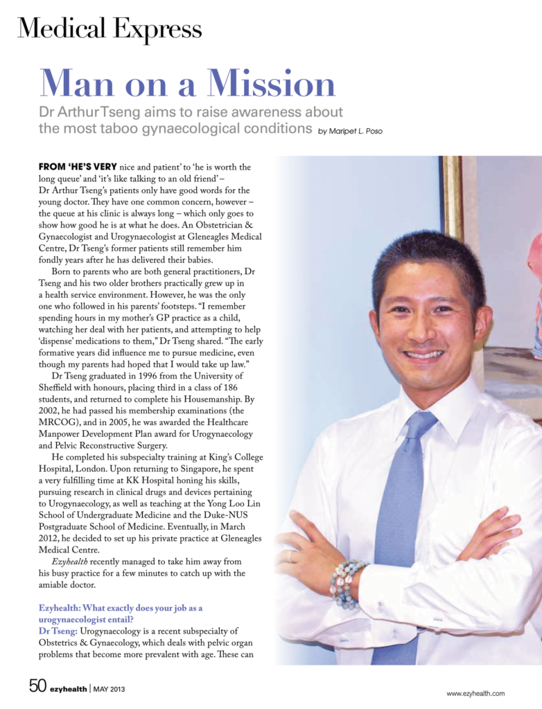
Dr Tseng's expertise covers urinary incontinence and pelvic organ prolapse, apart from that, his particular interest lies in the holistic management of Overactive Bladder Syndrome and other functional bladder conditions. With many years of experience in this field, he is committed to providing patients


